Separation of arteries and veins in pulmonary CT images to support lung disease detection
Our colleague Christian Payer successfully finished his master thesis on on May 28th, 2015. The goal of his work was to separate and analyze arteries and veins in thoracic computed tomography images. The proposed algorithm performs this task without manual intervention and is based on two integer programs. The results of the algorithm can be used to compare the morphology of arteries and veins. This information can support physicians to be able to perform an early diagnosis of lung diseases.
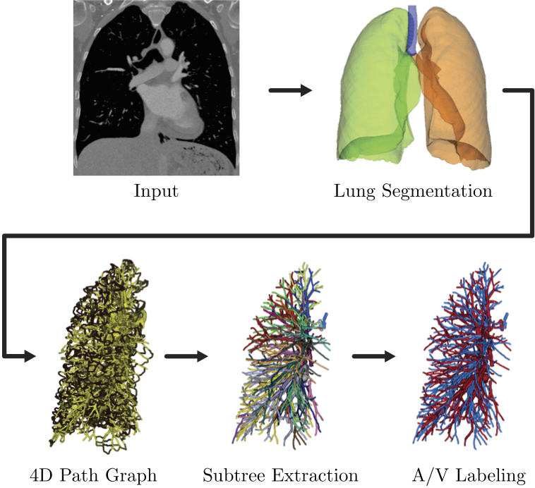
This is the flow chart of the proposed algorithm. The algorithm uses thoracic contrast-enhanced CT images as input. A lung segmentation is created and the A/V separation pipeline consisting of three steps, namely the 4D path graph, the subtree extraction, and the final A/V labeling, is applied to both sides of the lung independently.